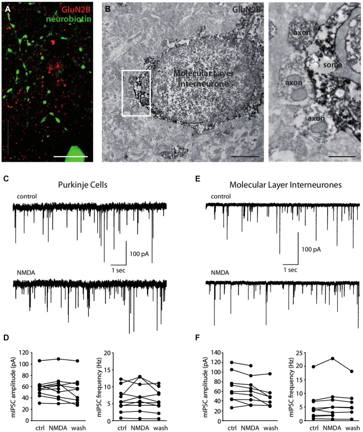
Figure 3.
Molecular Layer interneurons do not express functional axonal NMDA receptors. (A) Immunostaining showing the absence of GluN2B (red) colocalization with a neurobiotin-filled MLI axon (green). Sum of 56 optical slices in a stack. Scale bar = 10 μm. (B) Immunostaining for GluN2B showing GluN2B-stained putative inhibitory synapses (axon) on a GluN2B-stained MLI soma. Synapses are extended in the right panel. Scale bars: left panel 2 μm, right panel 0.5 μm. (C–F) Miniature IPSCs recorded in PCs (C,D) or in MLIs (E,F) in control conditions or in the presence of 15 μM (C,D) or 30 μM (E,F) NMDA. (C,E) Representative recordings. (D,F) Amplitude and frequency of mIPSCs for each cell before (ctrl), during (NMDA) or after (wash) bath-application of NMDA.

