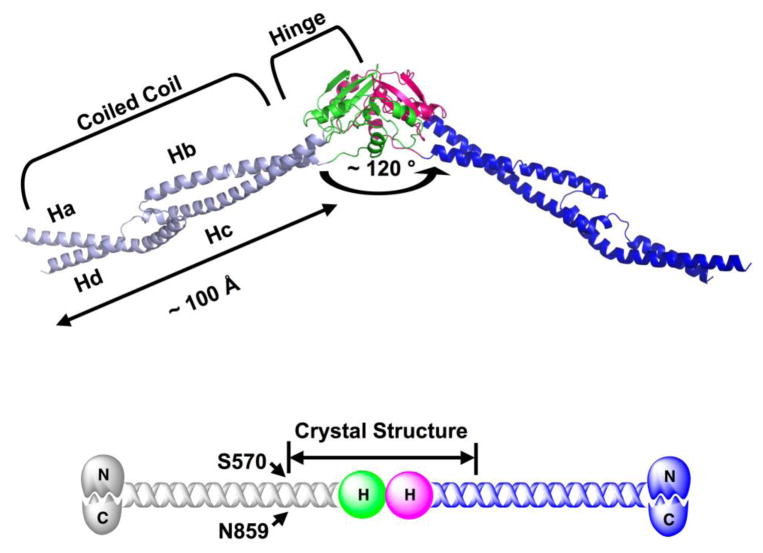
Fig 1.
Overall structure of MukB-D (566–863). Each MukB-D monomer contains the complete hinge domain (666–779) and two coiled coil strands (572–665 and 780–854). The angle of the V-shaped dimer is ~ 120°. The length of coiled coil domain in each monomer is ~ 100 Å. The hinge domains of two monomers are colored green and magenta respectively; the coiled coil domains for the same monomers are colored light blue and blue respectively. All figures involving crystal structures were prepared with PyMol (DeLano Scientific LLC). Experimental details for protein crystallization and structure determination are included in the supplementary material.

