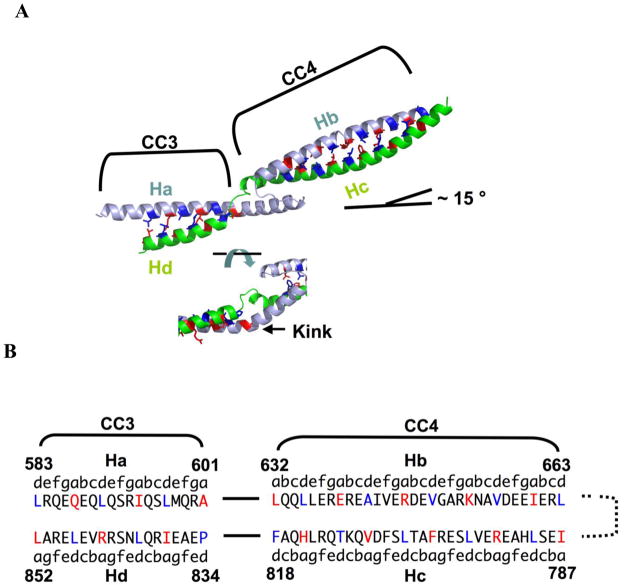
Fig 4.
Register analysis of coiled coil domain in E. coli MukB. (A) Close-up view of the coiled coil segment highlighting inter-strand packing (top) and the coiled coil discontinuity (bottom). The N- and C-terminal helical strands within the same monomer are colored light blue and green respectively. The coiled coil regions were assigned by the computer algorithm SOCKET 41 (packing cut-off = 7.4 Å). The two regions of the coiled coil are denoted CC3 (583–601 and 834–852) and CC4 (632–663 and 787–818). The side chains of residues at a (red) and d (dark blue) positions are shown as sticks. (B) Sequence of coiled coil strands highlighting residues involved in inter-strand packing interactions.

