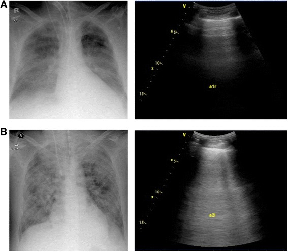
Figure 2.

Chest radiographs (left) and corresponding ultrasound screenshots (right) of two study patients. (A) Dry lung with a normal extravascular lung water index (EVLWI) and predominant A lines. (B) Severe, non-cardiac pulmonary edema with a high EVLWI and confluent B lines.
