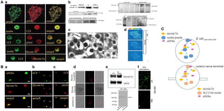
Figure 1.
(A) (a) Imaging studies of colocalization of nNOS, LC8, and myosin Va with insulin granules in pancreatic islets. Upper panel, pancreatic islets stained for insulin colabel with neuronal nitric oxide synthase (nNOS). Isolated islets colabel for insulin and LC8 (middle panel), nNOS and LC8 (third panel), insulin and myosin Va (fourth panel), and LC8 and myosin Va (bottom panel). (b) nNOS immunoblots in normal and diseased islets. Islets and INS1 cell line label for ~160 kDa nNOS band. Lower panels on the left show increased nNOS bands on the western blots obtained from fa/fa Zucker obese rats and obese human individuals, models of insulin hypersecretor phenotypes. The right panels show that nNOS exists as a dimer, revealed by cold SDS-PAGE. Dimer/monomer ratios are raised in the hypersecretor phenotypes. (c) Electron micrographs of insulin LDCVs (secretory granules). (a,e) Electron micrographs showing immune particles representing insulin and nNOS. In (e), note nNOS on the membrane of the LDCV. (g,h) Of the electron micrographs show nNOS-LC8 in the core and membrane of insulin LDCVs. (d) Ionomycin and l-arginine enhances NO production in INS1 cell lines, imaged by loaded diaminofluorescein. [Figures modified with permission from Lajoix et al. (17), Mezghenna et al. (16) and Smukler et al. (22).] (B) (a–c) Imaging studies of colocalization of nNOS, LC8, and myosin Va in isolated enteric synaptosomes. (d) Proximity ligation assay (PLA) shows blobs of protein interactions of nNOS, LC8, and myosin Va in isolated enteric synaptosomes. (e) Upper panel shows co-immunoprecipitation of nNOS-myosin Va in mice stomach lysate; lower panel shows intact nNOSα in whole varicosities of wild type and DBA/2J dilute mice, but absence of membrane bound nNOSα in DBA/2J, indicating the potential role of myosin Va in membrane transposition of nNOS. (f) KCl stimulation of plated varicosities shows significantly reduced DAF-NO signal in enteric synaptosomes obtained from DBA/2J mice, in comparison to C57BL/6J mice. [Figures modified with permission from Chaudhury et al. (11, 12).] (C) Cartoon depicting similarity in mechanisms of transcytosis of insulin and nNOS by myosin Va in beta cells and enteric synaptosomes. Note the similarity of organization of non-vesicular nNOS with either SLC17A9 purinergic vesicles within nerve terminals or insulin granules in beta cells of islets of pancreas. Genomic inhibition of myosin Va may be a potential initial upstream pathophysiologic mechanism contributing to both progression of diabetes by impairing insulin exocytosis, as well as causing multiorgan dysfunction, for example, reduction of inhibitory nitrergic neuromuscular transmission in the gut. Arrows are shown to indicate directionality of movements.

