
Figure 3.
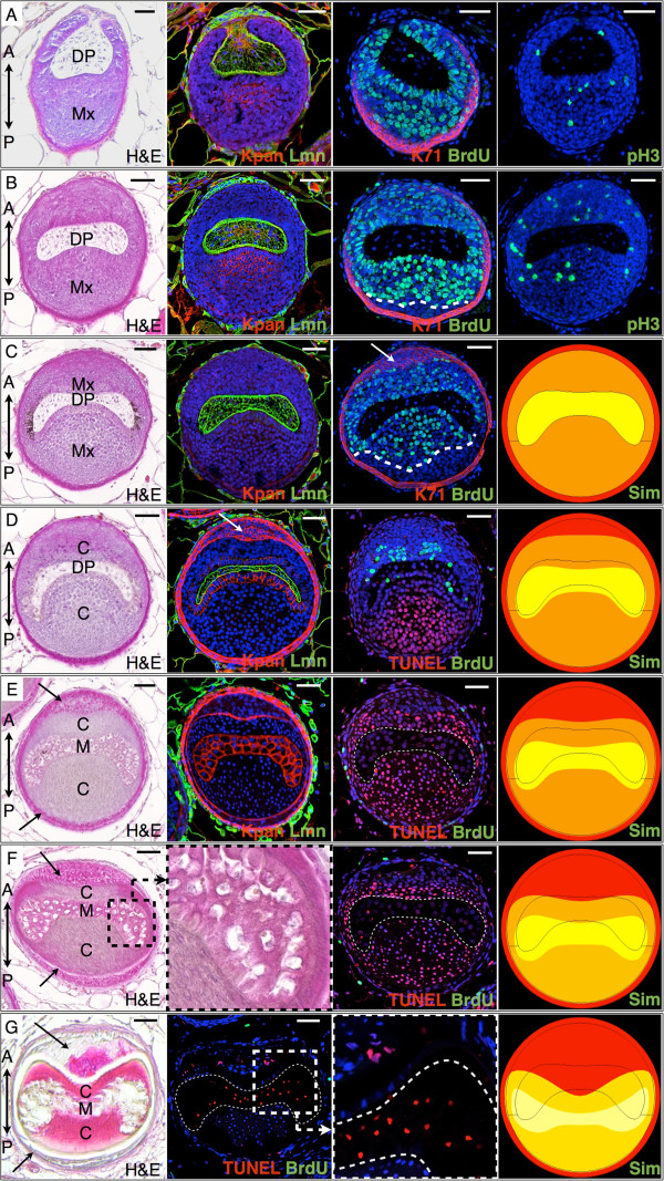
Observed and simulated cell proliferation and cell death along a spine follicle. Transverse sections (28-day-old Acomys) are taken along the spine long axis, from the basis of the follicle (rows A-C), to the DP-medulla transition (rows D-E), to the collapse of the keratinized medulla (rows F-G). H&E, Hematoxylin and Eosin staining. Immunostaining: Kpan, Pan-Keratin (red); Lmn, Laminin (green); BrdU, 5-bromo-2'-deoxyuridine (green); K71, keratin 71 (red); pH3, phospho-Histone H3 (green); TUNEL (red), Hoechst (blue). Arrows: IRS, DP: dermal papilla, Mx: matrix, C: cortex, M: medulla. Simulated transverse sections (Sim) are snapshots of the Supplementary Movie 2 showing time evolution of a simulated follicle. Scale bars: 50 μm.