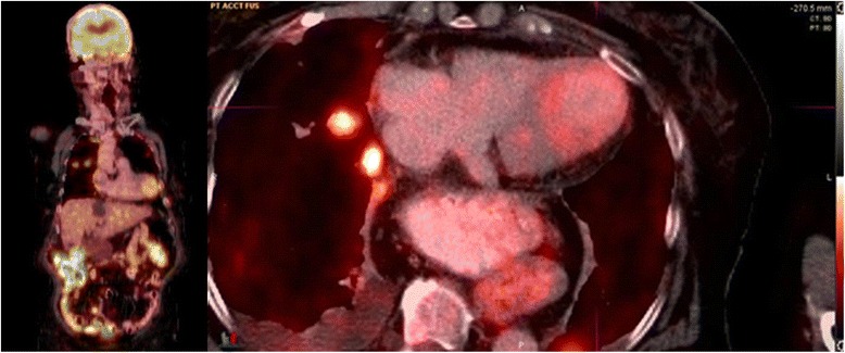
Figure 2.

Coronal (left) and axial (right) PET/CT showing a focus of mildly increased FDG uptake corresponding to ballooning of the left ventricular apex. Additional findings at the axial level through the chest include FDG-avid metastatic lung nodules in the right upper lobe, small right pleural effusion and a large hiatal hernia.
