
Figure 1.
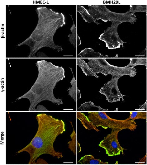
Localization of β- and γ-actin in vascular endothelial cells. Representative photographs of HMEC-1 (left) and BMH29L (right) endothelial cells stained with β-actin (top) and γ-actin (middle) antibodies. The merged photographs (bottom) show β-actin in green, γ-actin in red and DNA (DAPI) in blue. Scale bar, 20 μm.