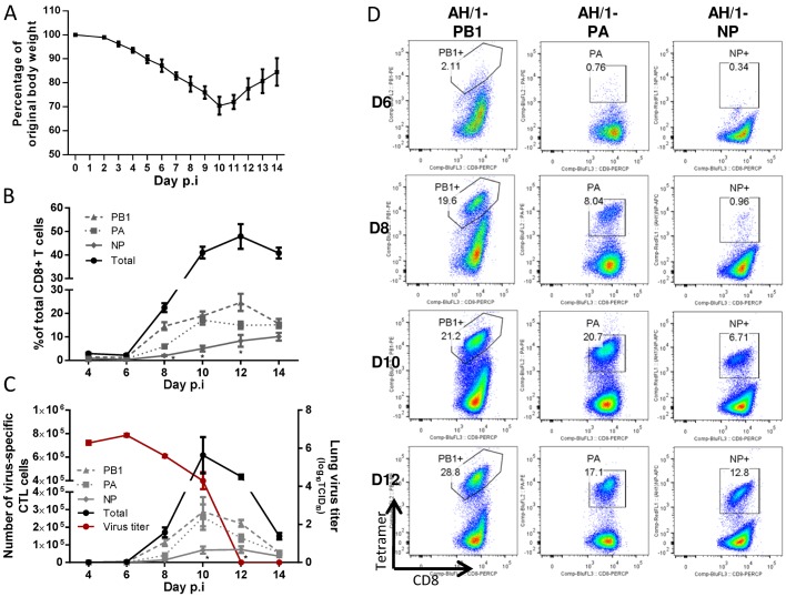
Figure 4. The disease course and primary CTL responses in naive mice after infection with the H7N9 IAV.
Naïve mice were infected with 1 MLD50 (103.5 TCID50) of the H7N9 virus. (A) Body weight change, (B) the proportion and (C, left Y axis) number of each epitope-specific CTL population in the BAL, and (C, right Y axis) the lung virus titer. Data sets represent mean ± SEM, n = 4–5 at each time point. * p<0.05, Tukey’s test, NP versus the other two epitopes. (D) Representative flow cytometry plots for each tetramer-specific CTL response in the BAL. The tetramers used were specific to the KbPB1703, DbPA224, and DbNP366 variants of the H7N9 virus.

