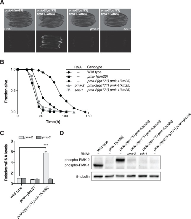
Fig 3. A deletion in the 3’UTR of pmk-2 confers an increase in PMK-2 expression that can substitute for PMK-1 activity in the intestine.
(A-B) Phenotypic analysis of the pmk-2(qd171) pmk-1(km25) mutant. Bright field and fluorescence microscopy images (A) and P. aeruginosa pathogenesis assays (B) of worms treated with RNAi as indicated for two generations. (C) qRT-PCR analysis of pmk-2 and pmk-3 mRNA levels in L4 larval stage animals. Levels of pmk-2 and pmk-3 mRNA are normalized to the levels of snb-1 mRNA. Values plotted are the fold changes relative to wild type. Shown is the mean ± SEM (n = 4 independent biological replicates, *** P<0.001, two-way ANOVA with Bonferroni post-test). (D) Immunoblot analysis of lysates from RNAi-treated mixed stage animals using an antibody recognizing the doubly phosphorylated TGY motif of activated PMK-1 and PMK-2 p38 MAPKs and an antibody that recognizes β-tubulin. (A-D) All strains carry the agIs219[PT24B8.5::GFP] transgene.

