
Fig 5. Autosomal synapsis in H3f3b heterozygous spermatocytes.
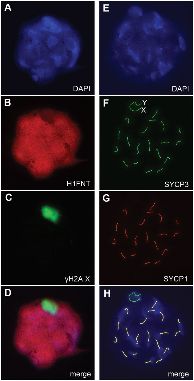
H3f3b -/+ male, 3 wk old. Spermatocyte 1: (A) DNA, DAPI stained, (B) mid-late pachytene spermatocytes are H1FNT-positive, (C) γH2A.X is localized to the XY-body. No γH2A.X staining evident in autosomes, indicative of normal synapsis, (D) merge of the three upper panels. Spermatocyte 2: (E) DNA, DAPI stained, (F) SYCP3 marks the chromosomal axial elements. By pachytene, these have coalesced into rod-like structures, (G) SYCP1 marks synapsed chromatin. The X and Y chromosomes are unsynapsed, therefore are negative for this marker, (H) merge of the three upper panels shows that SYCP3 and SYCP1 are coincident, indicating complete synapsis.