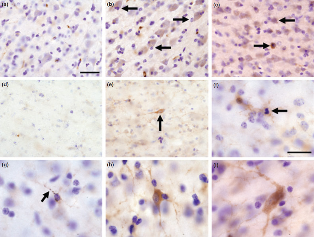
Fig. 1.
Representative images of APLP1 immunohistochemistry in the frontal cortex of control (a and d) and Mn-exposed animals (b, c, and e–i). Control animals exhibited faint APLP1 staining in cortical neurons (a) and in the white matter (d). In the frontal cortex of Mn exposed animals, there were darkly stained cells whose morphology resembles pyramidal cells (arrows in b) and cortical interneurons (arrow in c and cell in panel h). White matter interstitial neurons with fusiform morphology (arrow in e) and polymorphous morphology (i) also expressed increased APLP1 labeling. An overall increase in APLP1 was detected in glial cell processes in the white matter (e–g). The increase is apparent by comparing panel (d) (control) and panel (e) (Mn-exposed) white matter. In panel (f), there are two glial cells that expressed increased APLP1 immunoreactivity and one of the glial cells appears to have a condensed nucleus resembling apoptosis (arrow in f). In panel (g), there is a glial cell whose processes are APLP1 positive with a beaded appearance as if it was undergoing degeneration (arrow in g). Scale bar: (a–e) 40 and (f–i) 20 µm.

