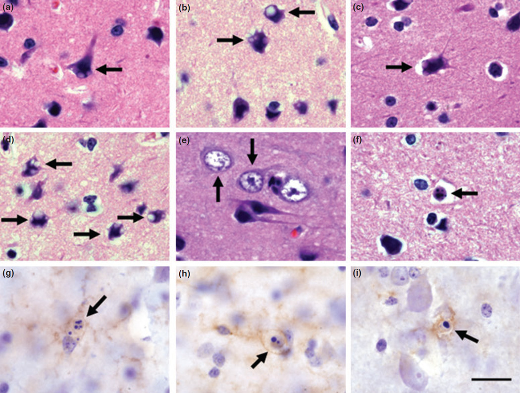
Fig. 4.
Morphological changes in the frontal cortex of Mn-exposed animals. Cortical cells of various morphological types displayed single or multiple intracytoplasmic vacuoles (arrows in a–d). We also observed cells in the frontal cortex of Mn-exposed animals with hyper-trophic nuclei and reduced cytoplasm (arrows in e). Cortical cells with apoptotic stigmata were present in Mn-exposed frontal cortex (arrows in f–i). Apoptotic cells could be seen with two or more spherical bodies (arrows in f–h) or with a single, dense, and spherical nuclear body (arrow in i). Cellular remnants of cells with apoptotic stigmata expressed APLP1 (g), hydroxynonenol (h), or p53 (i) immunolabeling. Panels (a–f) are hematoxylin and eosin staining and (g–i) are count-erstained with Nissl. Scale bar: (for all panels) 20 µm.

