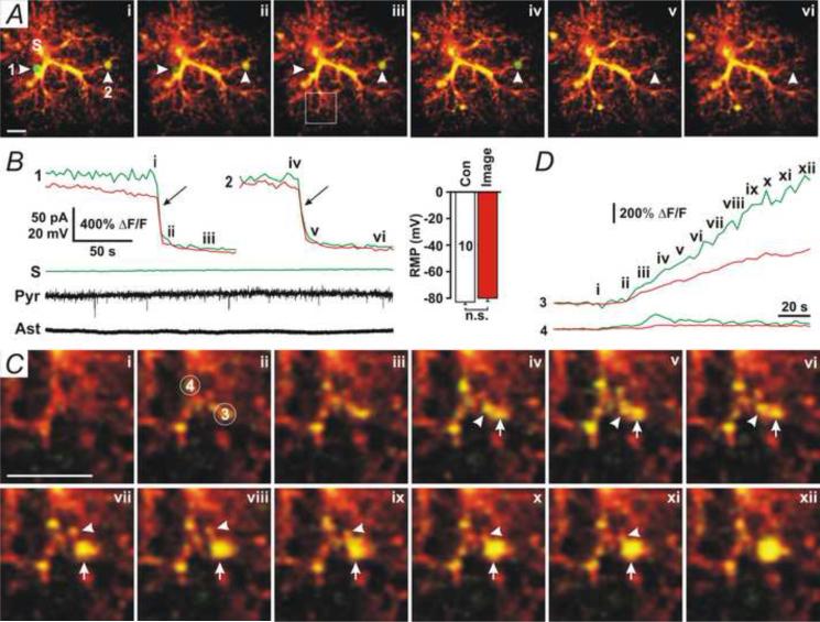
Fig. 3. High astrocytic [Ca2+] induces large vesicles.
(A) Two-photon microscopy images of an astrocyte that was patched with the pipette solution added with 100 nM [Ca2+], Fluo-4 potassium (100 μM), and Alexa Fluor-594 (100 μM). Two large vesicles (1 and 2) were undergoing fusion (i-ii and iv-v). (B) The time course of Fluo-4 (Green) and Alexa Fluor-594 (Red) fluorescence from the two large vesicles (1 and 2) and the soma (S) indicated in A, showing the occurrence of fusion events (arrows). Whole-cell voltage-clamp recording in a nearby pyramidal neuron (Pyr) with apical dendrites passing through the domain of the patched astrocyte showed no fusion-associated SICs. Whole-cell current-clamp recording in the patched astrocyte (Ast) showed no changes in resting membrane potential (RMP). Representative data were chosen from 8 experiments. Right insert, the mean RMP of patched astrocytes during DIC imaging (Con) or fluorescence imaging (Image). (C) The enlarged image in the square area in A-iii. Alexa Fluor-594 and Fluo-4 fluorescence first increased (ii – iii, 3 and 4) and then a large vesicle (3) formed. Arrows indicate the large vesicle, and arrowheads indicate that two small Alexa Fluor 594- and Fluo4-positive compartments were merging into the large vesicle. (D) The time course for Fluo-4 (Green) and Alexa Fluor-594 (Red) fluorescence from the circles (3 and 4) indicated in C. Scale Bars in A and C, 5 μm.

