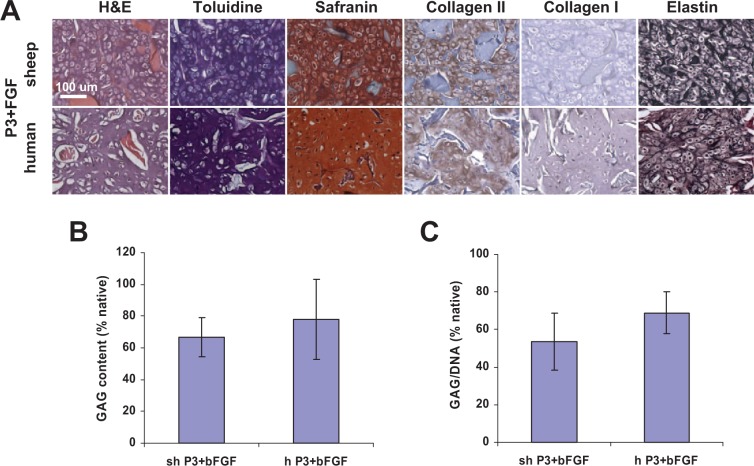
Figure 3.
Histological and biochemical assessment of elastic cartilage engineered from passaged P3 auricular chondrocytes expanded in the presence of bFGF after 6 weeks in nude mice. (A) Neocartilage formed from both sheep and human cells as evidenced by positive toluidine blue, safranin, collagen type II, and elastin stains. Elastin fibers were dense and strongly stained. Collagen scaffold fibers stained red on hematoxylin–eosin (H&E) stained sections. Scale bar: 100 µm. (B) GAG and (C) GAG/DNA ratio quantification confirmed histological results.

