
Figure 5.
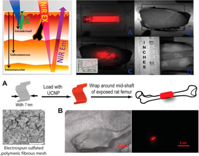
(top) left: the tissue depth of NIR and visible light. right: (a) UCPL bright-field image of a cuvette filled with a suspension of the core/shell nanoparticles, (b) bright-field image of a cuvette covered with pork tissue with a quarter coin stood aside showing its thickness, (c) merged UCPL/bright-field image of the cuvette covered with pork tissue, and (d) bright-field image of the pork tissue (side view). The inset in (c) shows the spectra obtained from the circled areas. (bottom) Polyethylenimine-coated NIRin-NIRout R-(NaYbF4:0.5%Tm3t)/CaF2 core/shell nanoparticles for imaging a synthetic periosteal mesh implanted around a rat femur. (a) UCNPs were loaded on a 7-mm-wide sulfated polymer mesh and wrapped around the mid shaft of a rat femur. Scale bar: 500 μm. (b) Bright-field image of the rat hind leg after closing muscle/skin by suture (left) and PL image (right) of the deeply embedded UCNP-stained synthetic mesh wrapped around the rat femur. Scale bar: 2 cm. (Reprinted with permission from ref (34). Copyright 2012 American Chemical Society.)