
Figure 6.
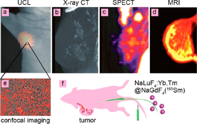
Four-model imaging of the focused tumor from the tumor-bearing nude mouse 1 h after intravenous injection of NaLuF4:Yb,Tm@NaGdF4(153Sm): (a) In vivo UCL-image, (b) X-ray CT image, (c) SPECT image, (d) MR image of tumor. (e) UCL confocal image of the paraffin section of tumor tissue. (f) Schematic illustration of tumor angiogenesis imaging using aLuF4:Yb,Tm@NaGdF4(153Sm) as the probe. (Reprinted with permission from ref (36). Copyright 2013 American Chemical Society.)