
Figure 7.
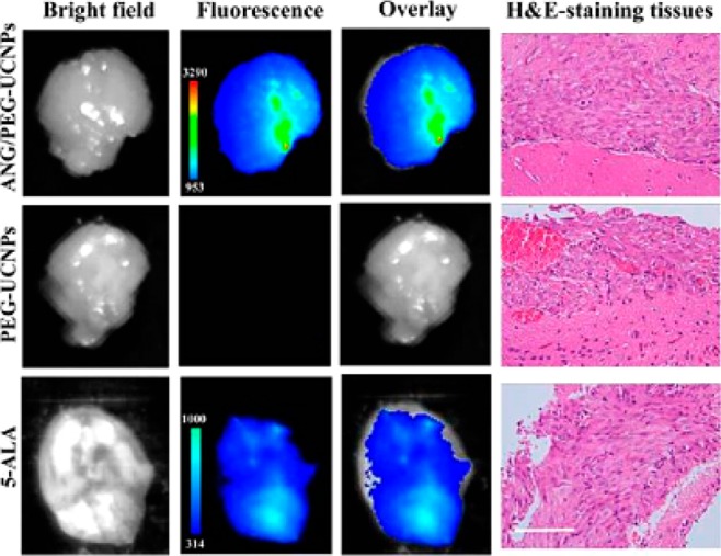
Ex vivo fluorescent images of glioblastoma-bearing brain in 1 h after the intravenous injection with ANG/PEG-UCNPs, PEG-UCNPs (excitation, 980 nm; emission, 800 nm), and 5-ALA (excitation, 470 nm; emission, 650 nm). All imaging experiments were performed under the same condition. H&E-staining of the tumor tissues from glioblastoma-bearing mice brain was used to demonstrate the existence of glionblastoma. Scale bar: 100 μm. (Reprinted with permission from ref (38). Copyright 2014 American Chemical Society.)