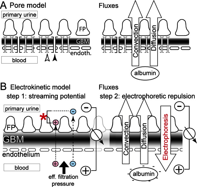
FIGURE 1 —

Schematic of the models for glomerular filtration. A. In the pore model, the glomerular filter is envisioned as an impermeable wall perforated by highly defined pores. Small pores (white arrow) allow the passage of water and small solutes, while rare large pores (black arrow) allow the passage of macromolecules. Two fluxes drive albumin across the barrier: convection and diffusion. Only the small size of the pores restricts albumin from passing. Proteinuria is understood as a pore-pathy, where pore diameter increases. B. In the electrokinetic model, a streaming potential (asterisk) is generated by the effective filtration pressure and slight differences in the passage of small ions (red, blue) (step 1). This electrical field acts on negatively charged albumin, preventing its passage across the barrier (step 2). Electrophoresis prevents the influx of albumin into the filter driven by convection and diffusion. FP = podocyte foot processes; GBM = glomerular basement membrane.
