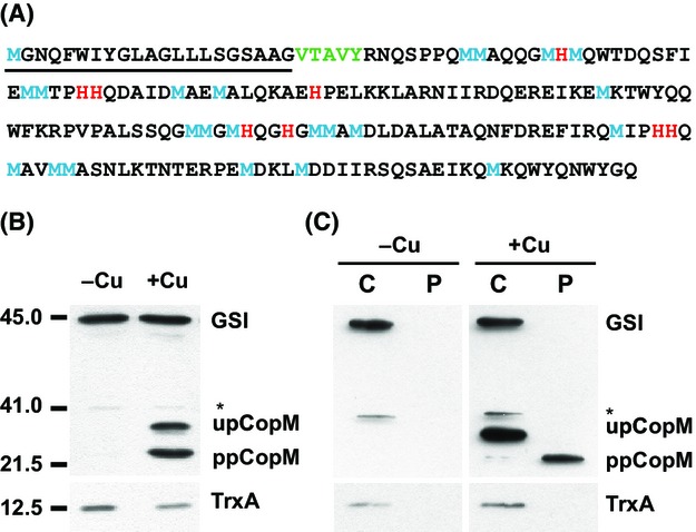
Figure 1.

CopM is a periplasmic protein induced by copper. (A) CopM amino acid sequence. The signal peptide sequence is underlined, the most likely cleavage site is shown in green, methionine and histidine residues are shown in blue and red, respectively. (B) Western blot analysis of CopM in the presence or absence of copper. WT cells were grown in BG11C-Cu medium to mid-log growth phase and exposed for 4 h to copper 1 μmol/L. Five micrograms of total protein from soluble extracts was separated by 15% SDS-PAGE and analyzed by western blot to detect CopM, thioredoxin A (TrxA), and glutamine synthetase type I (GSI). (C) Western blot analysis of CopM cellular localization. WT cells were grown in BG11C-Cu medium to mid-log growth phase and exposed for 4 h to copper 1 μmol/L. Five micrograms of both cytosolic (C) and periplasmic (P) protein from soluble extracts was separated by 15% SDS-PAGE and analyzed by western blot to detect CopM, TrxA, and GSI. upCopM, unprocessed protein; ppCopM, processed protein.
