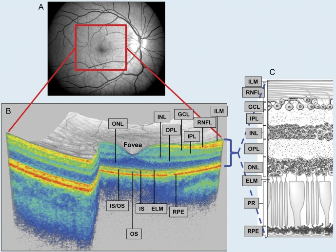
Figure 1. Eye of a healthy control subject.
(A) Photograph of fundus. (B) A 3-dimensional macular volume cube generated from the macular region (outlined in red in panel A). Optical coherence tomography segmentation allows determination of retinal layer thicknesses: macular retinal nerve fiber layer (RNFL), GCL + IPL (ganglion cell layer and inner plexiform layer), INL + OPL (inner nuclear layer and outer plexiform layer), and the outer nuclear layer (ONL), which includes the inner and outer photoreceptor segments. Individual retinal layers are readily discernible, but the GCL cannot easily be distinguished from the IPL. (C) Cellular composition of retinal layers. ELM = external limiting membrane; ILM = inner limiting membrane; IS = inner segment; OS = outer segment; PR = photoreceptors; RPE = retinal pigment epithelium. Copyright © 2013 American Medical Association. All rights reserved.

