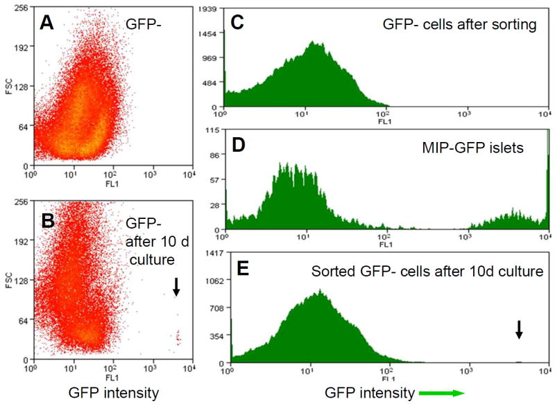
Fig. 3.

MoFlo sort. a, c MoFlo sort of dispersed GFP-negative samples after COPAS sort showed that there were no GFP cells in this population. b, e GFP-positive differentiated cells in the GFP-negative population 10 days after the COPAS sort. Arrow, new GFP-positive cells. d The MIP-GFP islet sample from COPAS serves as a positive control. FL1,conventionally used for green channel; FSC forward scatter which indicates cell volume
