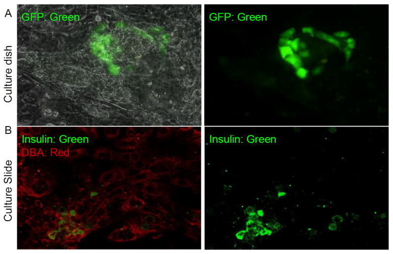
Fig. 4.

GFP/insulin protein is expressed in cystic structures of epithelial monolayer from GFP- population 10 days after COPAS sort. a A cluster of GFP-positive cells (green) seen within monolayer (overlaid on phase image on left and just green channel on right panel. b Co-staining for insulin (green) and the ductal marker DBA lectin (red) suggests that new insulin-positive cells are derived from duct cells in vitro (merged with both channela on left and just green for insulin on the right panel).
