Figure 1.
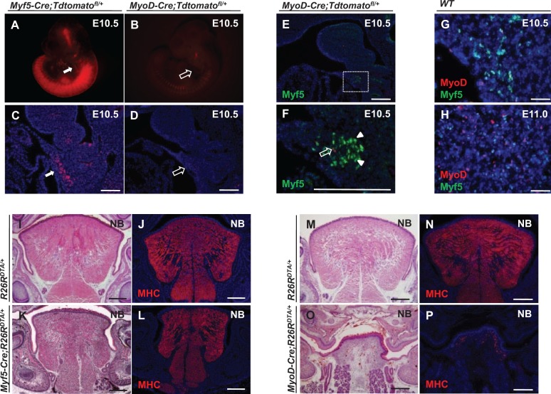
Myf5- and MyoD-expressing cells are distinct subpopulations during tongue myogenesis. (A–D) Detection of Myf5- and MyoD-derived cells in E10.5 Myf5-Cre;Tdtomatofl/+ (A, C) and MyoD-Cre;Tdtomatofl/+ (B, D) reporter mice, respectively. Myf5-derived cells are detectable in the hypoglossal cord (white arrows), whereas few MyoD-derived cells are detectable (open arrows). (E, F) Myf5 immunostaining (green, arrowheads) in the hypoglossal cord region of E10.5 MyoD-Cre;Tdtomatofl/+ reporter mice (MyoD-derived myogenic cells indicated by red, open arrow). Boxed area (E) is shown magnified (F). (G, H) Double immunostaining of MyoD (red) and Myf5 (green) in the hypoglossal cord at E10.5 (G) and the tongue bud at E11.0 (H) of wild-type mice. (I–L) Hematoxylin and eosin staining (I, K) and myosin heavy chain (MHC) immunostaining (J, L) of tongues from newborn (NB) Myf5-Cre;R26RDTA/+ and R26RDTA/+ control mice. (M–P) Hematoxylin and eosin staining (M, O) and MHC immunostaining (N, P) of tongues from NB MyoD-Cre;R26RDTA/+ and R26RDTA/+ control mice. Scale bars, 200 µm.

