Figure 5.
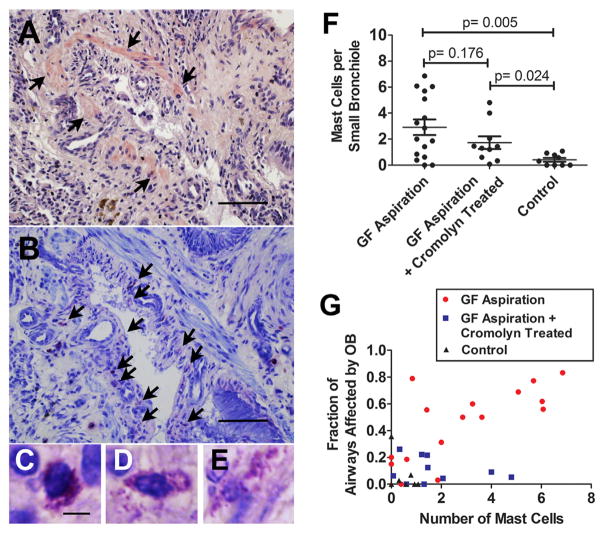
Mast cells in the small bronchioles. Serial sections using hematoxylin and eosin stain (A) and toluidine blue stain (B) show an OB lesion in a pulmonary allograft exposed to 8 weekly aspirations of gastric fluid (GF). In (A), arrows denote the smooth muscle of the bronchiole, and in (B) arrows indicate mast cells associated with the bronchiole. Different stages of degranulation of mast cells were noted in the bronchiole using toluidine blue staining (C–E). The number of mast cells per small bronchiole increased significantly in animals aspirated with GF, regardless of cromolyn treatment (GF aspiration vs control: p = 0.005; GF aspiration plus cromolyn vs control: p = 0.024). There was no significant difference between the number of mast cells per bronchiole in animals with and without treatment with cromolyn (GF aspiration vs GF aspiration plus cromolyn: p = 0.176) (F). A positive correlation was observed between OB and the number of mast cells in allografts from the GF aspiration group (r = 0.709, p = 0.002), but not in allografts from the GF aspiration plus cromolyn group (r = −0.225, p = 0.532) or the control group (r = −0.297, p = 0.439) (G). Bar = 200 μm in (A) and (B) and 5 μm in (C)–(E).

