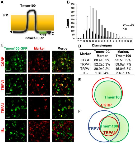
Figure 1.
Tmem100 is expressed in TRPA1/TRPV1 positive peptidergic DRG neurons. (A) Postulated structure of Tmem100. Tmem100 is a two-transmembrane protein with a putative TRPA1 binding site (KRR) at its C-terminus. Both the N- and C-termini are intracellular whereas the loop region is extracellular. “PM”: plasma membrane. (B) Tmem100-expressing cells comprise 24% of L4-L6 DRG neurons. They are predominantly small-diameter, but medium- to large-diameter neurons are also present. The average diameter of Tmem100-expressing neurons is 15.7 μm, and the median is (A) 14.3 μm (DRG from 3 mice). (C) Double staining of Tmem100-GFP with other DRG markers. Bar: 50 μm. (D) Quantification of co-expression of Tmem100 and other DRG markers (DRG from 3 mice; data are presented as mean ± SEM). (E)(F) Diagrams showing the relationship of Tmem100 with other DRG markers. Tmem100 is a marker for the majority of CGRP+ DRG neurons (E); most TRPA1+ DRG neurons express Tmem100 (F).

