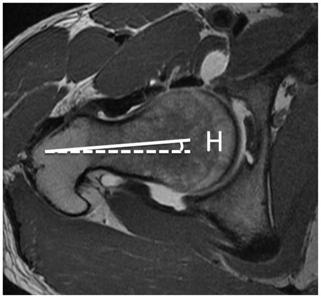
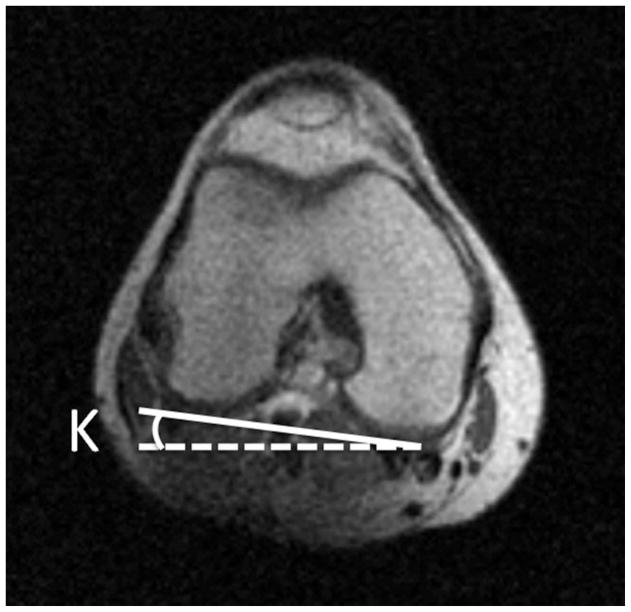
Figure 2.
(a) Axial T1-weighted image through the right femoral neck, with a line drawn along the axis of the femoral neck (solid line), and a horizontal line (dotted line). The angle between these lines is labeled angle H. (b) Axial image through the distal femur with a line drawn along the femoral condyles (solid line) and a horizontal line (dotted line). The angle between these lines is labeled angle K. If knee is internally rotated as in this example, the angle of antetorsion = H + K. If the knee is externally rotated the angle of antetorsion = H − K.


