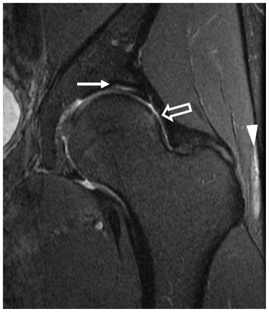
Figure 7.
Coronal fat-saturated T2-weighted image of the left hip in a patient with cam-type femoroacetabular impingement demonstrates delamination of the superior acetabular cartilage (arrow). Bone marrow edema is seen along the lateral femoral head-neck junction (open arrow). In addition there is non-specific edema between the iliotibial tract and greater trochanter (arrowhead).

