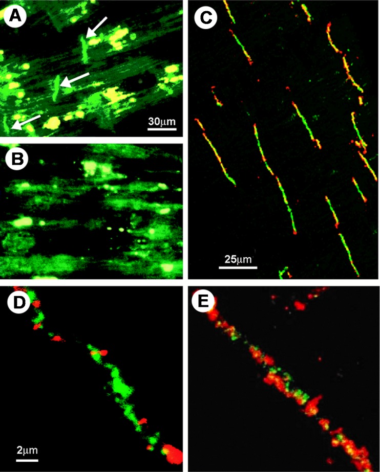
Fig. 3.
Immunohistochemical staining of P2X1 receptors in human left ventricle and co-localisation with connexin43. a P2X1 staining is shown (green, FITC) with arrows indicating the intercalated discs. Scale bar = 30 μm. b After pre-incubation of the antibody with epitope peptide, positive P2X1 staining at intercalated discs preferentially disappears, although some green and yellow auto-fluorescence remains (probably due to the highly fluorescent protein lipofuscin). Scale bar as in a. c Double labelling of P2X1 receptor and connexin43 in human left ventricle. Sections were double-labelled with anti-P2X1 (green, FITC) and anti-connexin43 (red, Cy3). The images were obtained by confocal microscopy. Both P2X1 and the gap junction protein connexin43 are localised in the intercalated discs. Note the variable degree of double labelling (yellow) in different discs. Scale bar 25 μm. d and e are higher magnification micrographs showing the variable amount of double labelling of P2X1 receptors and connexin43 in two different gap junctions. Scale bar in d and e 2 μm (reproduced from [286], with permission)

