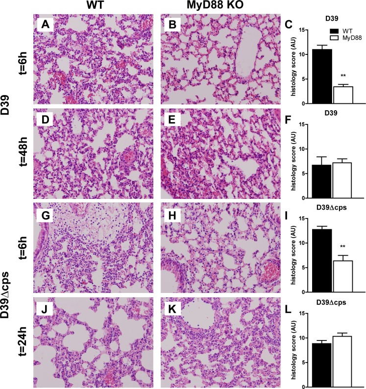
Fig 2. Lung pathology in WT and Myd88-/- mice during infection with capsulated and unencapsulated D39.
Representative lung sections obtained 6 or 48 h after induction of pneumococcal pneumonia with D39 in WT (A and D) and Myd88-/- mice (B and E) and 6 or 24 h after inoculation with D39Δcps in WT mice (G and J) and MyD88-/- mice (H and K). Haematoxylin and eosin stainings show different levels of inflammation. Original magnification 20x. Findings were quantified by histology scoring, as described in methods section for wild type (WT, black bars) and Myd88-/- mice (MyD88 KO, white bars). Data are mean ± SEM (N = 5–8 mice per group at each time point). **P<0.005 vs WT mice.

