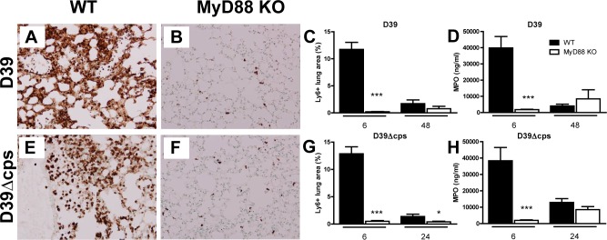
Fig 3. Reduced neutrophil infiltration in the lung in Myd88 -/- mice during infection with D39 or D39Δcps.

Representative Ly6-C/G stainings of lung sections obtained 6 h after induction of pneumococcal pneumonia with D39 in WT and Myd88-/- mice (A and B) and with D39Δcps in WT mice and Myd88-/- mice (E and F). Original magnification 20x. Percentage lung area with Ly6-C/G-positive neutrophils was quantified by digital image analysis as described in methods section for wild type (WT, black bars) and Myd88-/- mice (MyD88 KO, white bars)(C and G). Levels of MPO in lung homogenates of wild type (WT, black bars) and Myd88-/- mice (MyD88 KO, white bars) inoculated with D39 at 6 and 48 hours after challenge, or inoculated with D39Δcps (D and H) at 6 and 24 hours after challenge were determined by ELISA. Data are mean ± SEM (N = 5–8 mice per group at each time point). *P<0.05, ***P<0.0005 vs WT mice.
