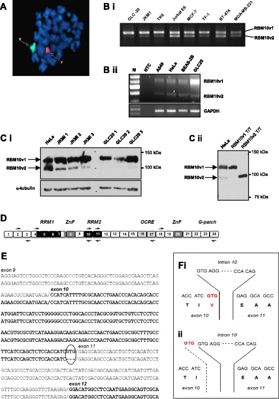
Figure 1.

Alternative splicing of RBM10. (A) FISH analysis of GLC20 cells, with painted X and Y chromosomes, demonstrating the presence of only one X chromosome. (B) RBM10v1 and RBM10v2 RNA expression in various cell lines, including GLC20. Representative raw RT-PCR data using RBM10 exon 4 spanning primers. Bi: RBM10F with RBM10RS primers. Bii: RBM10F with RBM10v1/v2R primers. M: 100 bp DNA ladder (FroggaBio Inc., Toronto, Canada). NTC: no template control. (C) Protein expression by Western blot. Ci shows RBM10 expression in whole cell lysates from three cell lines, including GLC20. The numbers 1, 2 and 3 after JKM1 and GLC20 delineate cells from three biological replicates. Cii includes control HeLa protein and in vitro translated RBM10v1 and RBM10v2 protein, to confirm the location of RBM10v2 is the cell line extracts. (D) Cartoon of full-length RBM10v1 mRNA, not drawn to scale. Boxes represent exons. Left and right black arrows represent primer placement for sequencing. Approximate positioning of consensus functional motifs is indicated by text and differential shading. (E) Alignment of the two GLC20 RBM10v1 isoform sequences. Circled area indicates the region that differs between the two RBM10v1 isoforms. (F) Nucleotide and amino acid sequences of the RBM10v1 exon10/intron 10/exon 11 donor and acceptor sites for (i) RBM10v1(V354), and (ii) RBM10v1(V354del).
