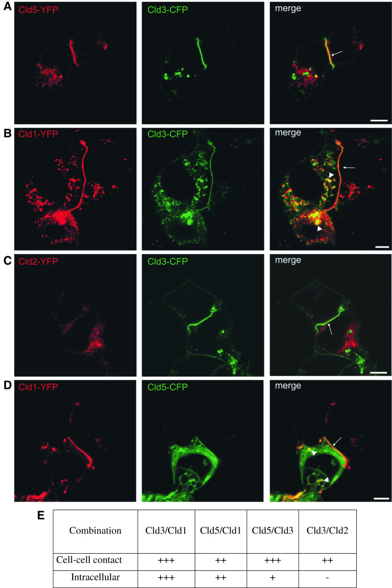
Fig. 4.
Differential colocalization of coexpressed claudin subtypes at cell–cell contacts and intracellular compartments. Pairs of Cld1, -2, -3, and -5 were coexpressed in HEK cells and the subcellular distribution analyzed by confocal microscopy. At contacts between two Cld-expressing cells (arrows) colocalization of Cld3/Cld5 (a), Cld3/Cld1 (b), Cld5/Cld1 (d), and partly Cld3/Cld2 (c) was observed. Strong colocalization in intracellular compartments (arrow heads) was found for Cld3/Cld1 (b), partially for Cld5/Cld1 (d) but not for Cld3/Cld5 (a) and Cld3/Cld2 (c). Bar, 5 μm (e) Summary of the colocalization analysis.−, no; (+)/+, weak; ++/+++, strong colocalization

