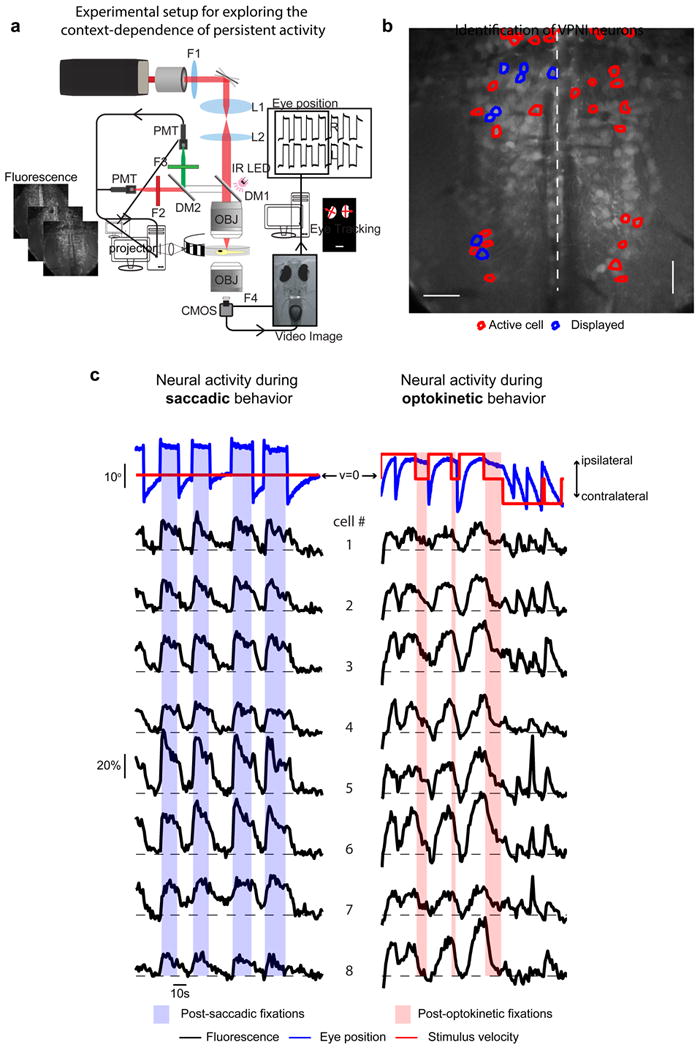
Figure 2. Experimental setup for exploring the context dependence of persistent activity.

(a) Schematic of experimental set-up used for synchronous two-photon calcium imaging, behavioral control, and eye-tracking (DM: dichroic mirror, F: filter, IR: infrared, L: lens, PMT: photomultiplier tube, OBJ: objective, CMOS: CMOS camera). (b) Average image of one plane in the caudal hindbrain where VPNI neurons were located. The Mauthner axons are visible on either side of the midline. Outlines indicate cells with eye position correlated activity; blue outlines indicate those that are plotted in (c). (c) Eye position and stimulus velocity (top), and fluorescence time series of individual cells (bottom) during saccadic and optokinetic eye movements. Dashed lines indicate the baseline level of fluorescence for each cell. Colored bars indicate post-stimulus fixation regions where gaze stability is dependent on persistent firing generated within the VPNI.
