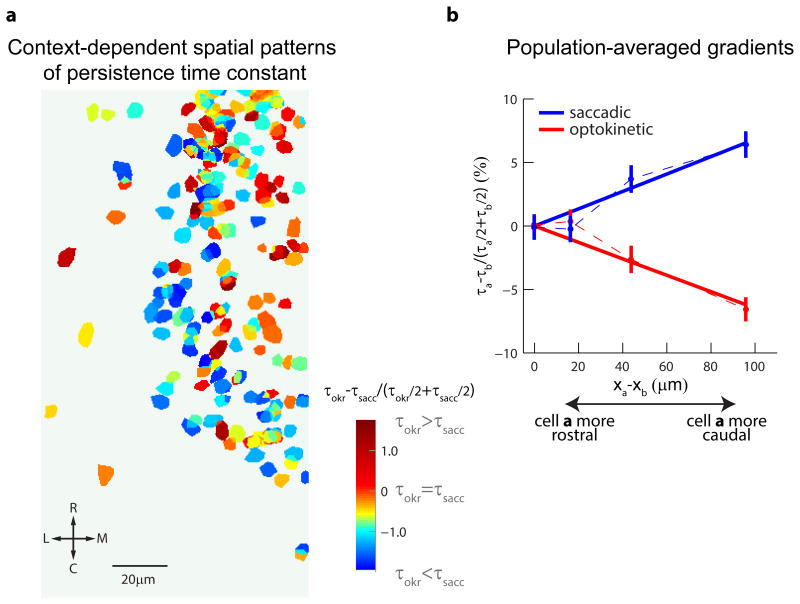
Figure 5. Context-dependent reversal in spatial gradients of persistent firing.
(a) Map of VPNI neurons from all animals aligned to the rhombomere 6/7 border (Figure S4). Neurons are color-coded according to the fractional difference between their time constant during post-optokinetic and post-saccadic fixations , with red indicating longer time constants during post-optokinetic fixations. (b) For the entire population, gradients in persistent firing rate along the rostro-caudal direction assessed by measuring pairwise differences in time constant, normalized by the pairwise mean time constant. Confidence intervals indicate the standard error of the estimate of the mean.

