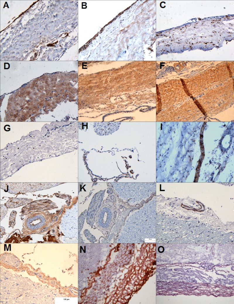
Figure 2.

Immunohistochemical analysis of the porcine meninges. The meninges composed of the dura mater (A–G), the arachnoid mater (H–I) and the pia mater (J–M) in formalin fixed tissue. The porcine spinal cord was cryosectioned and the porcine dura mater (N–O) was stained. The tissue sections were labeled for: (A) von Willebrand factor, (B and K) fibronectin, (C and J) vimentin, (D) laminin, (E) collagen I, (F and M) collagen II, (G, H, and L) integrin 1β, (I) Desmoplakin, (N) collagen III, (O) E-cadherin. Images were captured at ×200 magnification. [Color figure can be viewed in the online issue, which is available at wileyonlinelibrary.com.]
