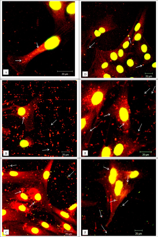
Figure 5.

Representative confocal microscopy images of cells exposed to green fluorescent nano-particles (40 nm in diameter). (A–C) Dural epithelial cells. (D–F) Dural fibroblasts. (A, D) Cells exposed for 1 day. (B, E) Cells exposed for 2 days. (C, F) Cells exposed for 3 days. White arrows indicate particles internalised whereas white dashed arrows indicate particles outside from cells. After day 2 there is evidence of particle agglomeration. The actin filaments were stained with rhodamine phalloidin and the cell nuclei with sytox green. All the images were taken at 630× magnification. In total 300 cells were imaged (three replicates × 100) at each time point. [Color figure can be viewed in the online issue, which is available at wileyonlinelibrary.com.]
