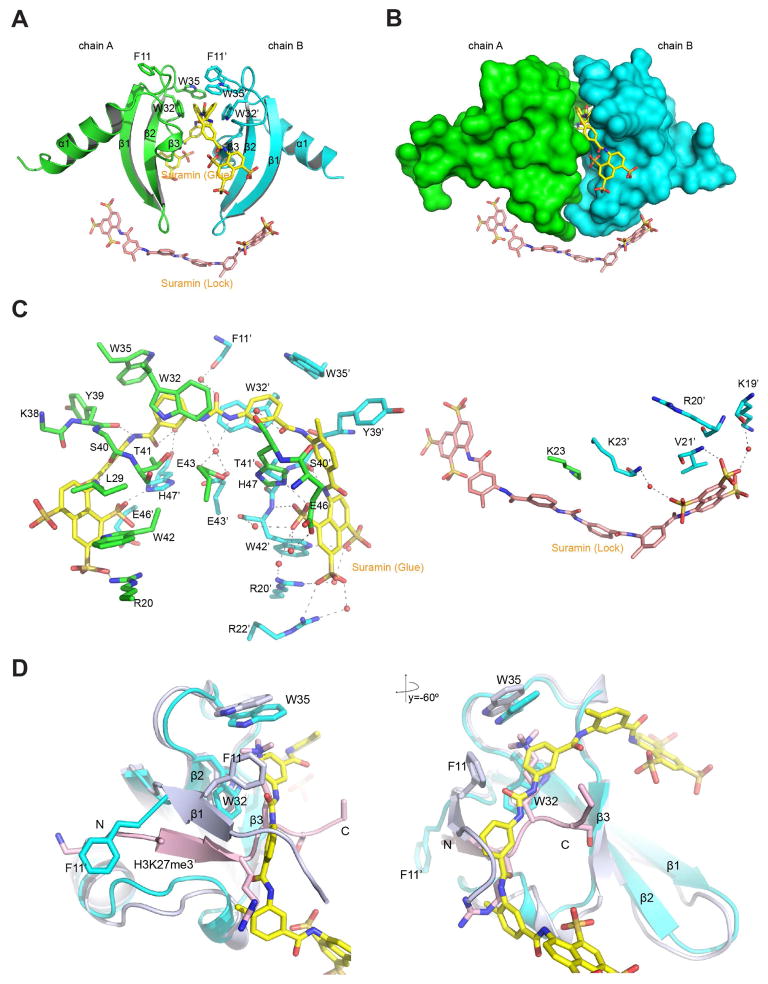
Figure 3. Structural analysis of Suramin binding to CBX7ChD.
(A) Crystal structure of a CBX7ChD dimer bound to suramin-glue (yellow) and suramin-lock (salmon) molecules.
(B) Surface presentation of CBX7ChD bound to suramin.
(C) Detailed analysis of the interactions between CBX7ChD and suramin.
(D). Structural comparison of CBX7ChD bound to suramin or H3K27me3 peptide. Chain A and suramin-glue from the CBX7ChD-suramin complex and Chain AC from CBX7ChD-H3K27me3 complex were aligned.

