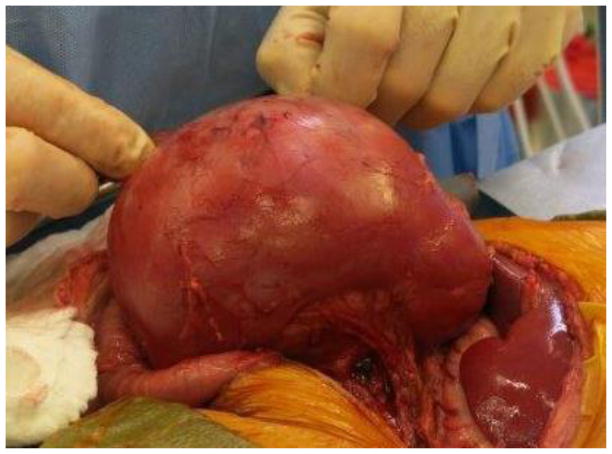
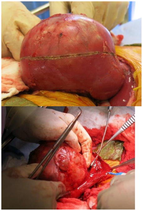
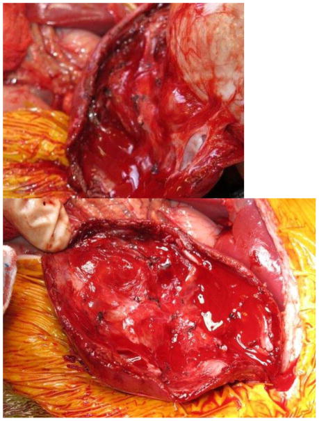
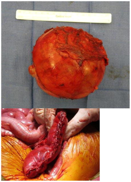
Figure 1. Nephron-sparing surgery.
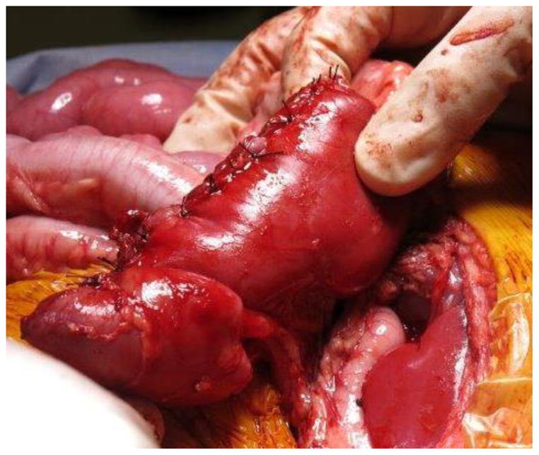
A. Kidney and tumor completely mobilized. B. Scoring of the capsule at the tumor-kidney interface. C. Dissection in the plane between tumor and kidney. D. An opening in the collecting system that requires closure with running monocryl suture. E. Cut surface of the remaining normal kidney parenchyma after ensuring hemostasis and closure of the collecting system. F. The resected specimen. G. Longitudinal folding over of the remaining normal kidney with reniform contour maintained with silk mattress sutures. H. View of the residual normal kidney at completion of nephron-sparing procedure.

