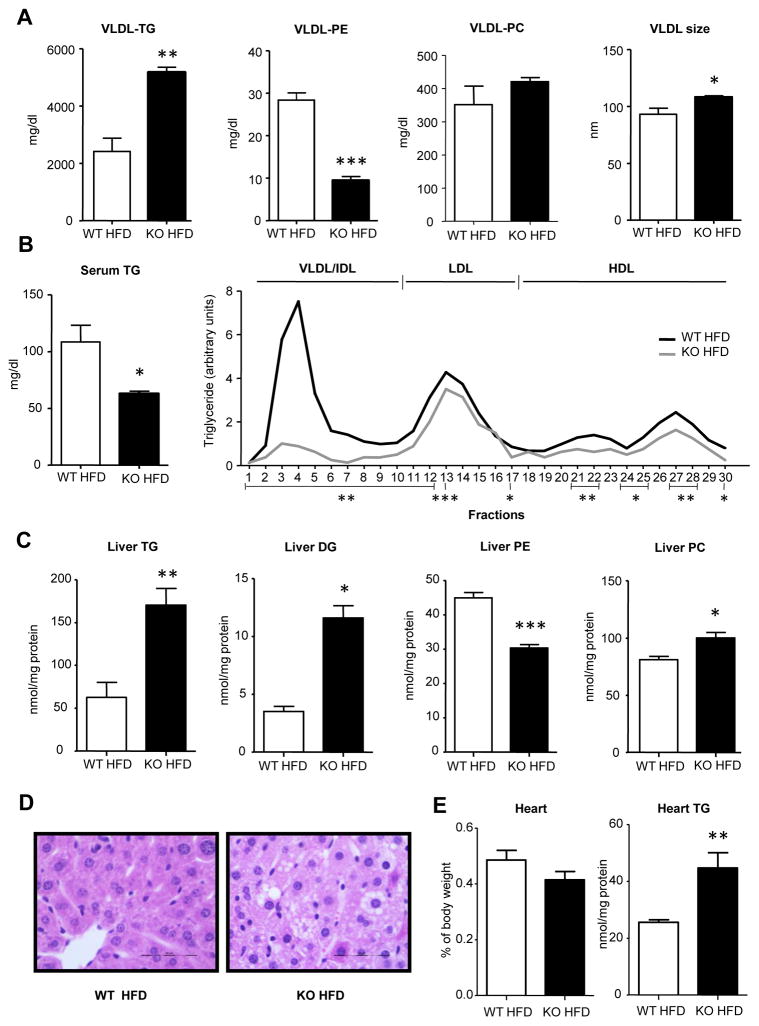
Figure 3. A HFD induces VLDL clearance and liver TG storage in GNMT-KO mice.
Wild type mice fed a high fat diet (WT HFD) and GNMT-KO mice fed a HFD (KO HFD) were fasted 2 hours. (A) Before 1 g/kg poloxamer (P-407) injection and 6 hours later, VLDL were isolated from serum and characterized for triglyceride (TG), phosphatidylethanolamine (PE) and phosphatidylcholine (PC) content and VLDL size. (B) TG levels in serum and in lipoprotein sub-fractions were measured. (C) Liver triglycerides (TG), diglycerides (DG), phosphatidylethanolamine (PE) and phosphatidylcholine (PC) levels from WT and KO mice fed the HFD were quantified. (D) Representative liver hematoxylin and eosin. (E) Percentage of heart weight. Heart TG levels were quantified after lipid extraction Values are mean±SEM of 4–5 animals per group. Statistical differences between KO and WT mice are denoted by *p<0.05; **p<0.01; ***p<0.001 (Student’s t test)

