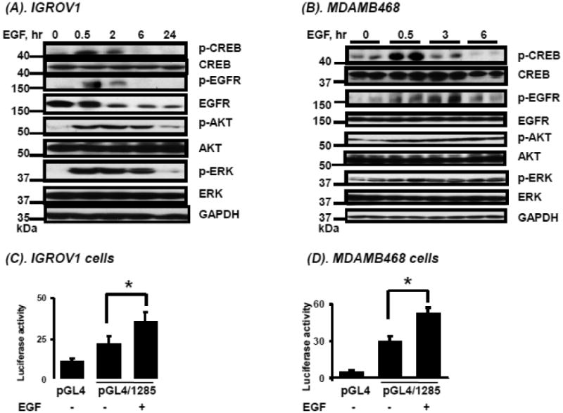
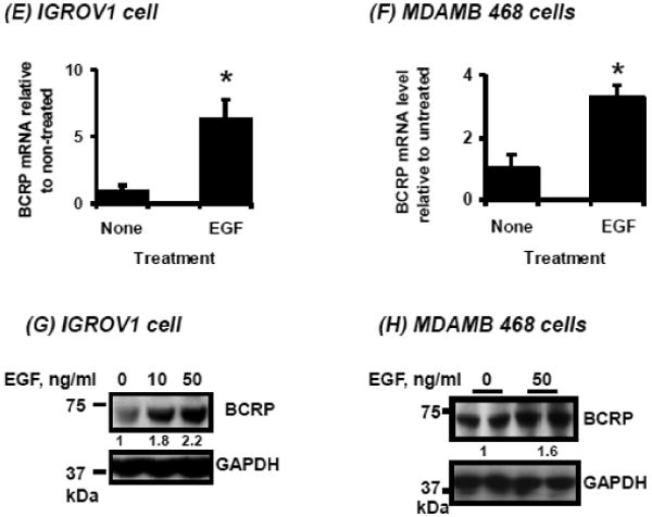
Figure 3.


Effects of EGF on pathways downstream of EGFR, CREB activation, and on BCRP promoter activity, mRNA and protein expression. A, B. Time-course of phosphorylated and unphosphorylated EGFR, AKT, ERK and CREB following EGF treatment (see Methods) in IGROV1 human ovarian carcinoma cells (A) and MDAMB468 human breast carcinoma cells (B) determined by Western blotting as described in “Methods.” C, D. The numbers on the left of the blot reflect the molecular size of marker proteins (kDa). Effects of EGF on activity of a BCRP promoter reporter construct transfected into human carcinoma cells. Cells were cultured, transfected with reporter constructs and exposed to EGF as described in Methods, then reporter activities were determined for IGROV1 ovarian (C) and MDAMB468 breast cancer cells (D). E, F. Effects of EGF on BCRP mRNA expression in human carcinoma cells. IGROV1 or MDAMB468 cells were treated with and without EGF as described in Methods, then total RNA was isolated and BCRP mRNA expression was were determined by real time qPCR for IGROV1 (E) and MDAMB468 cells (F). G, H. Effects of EGF on BCRP protein expression in human carcinoma cells. Cells were exposed to EGF as described in Methods, then harvested and BCRP expression was determined by Western blot in IGROV1 (G) and MDAMB468 cells (H). Western blots for GAPDH were used as a loading control. The data shown represent the mean and standard deviation of 3 different experiments, done on different days. Each individual assay was run in duplicate. *, significantly (P<0.05) different vs. untreated by student's t-test. Western blots shown represent one of three independent blots done on different days, with similar results obtained.
