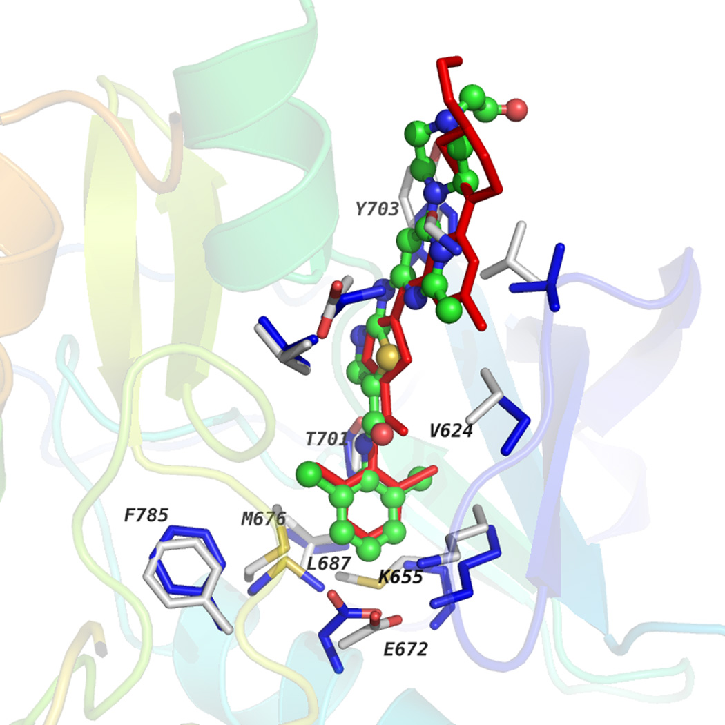
Figure 4.
Discoidin domain receptor 1 (DDR1) homology model complexed with dasatinib is shown along with the co-crystal structure of Abl kinase and dasatinib (PDB: 2GQG). Dasatinib docked into the DDR1-active state homology model is shown in the multicolored ball-and stick-model, whereas the native binding pose of dasatinib in 2GQG is shown in the red stick model. Residues in the binding pocket for DDR1 and Abl kinase are shown in gray or blue sticks, respectively. Residues that interact with ligands are labeled along with those that form the hydrophobic spine.

