
Figure 3.
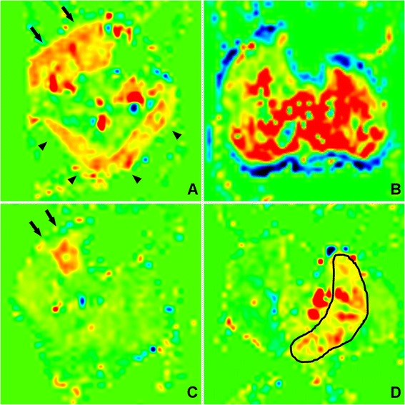
Representative perfusion maps of each study group demonstrate the difference in perfusion patterns with exercise (A, C) compared to cuff occlusion hyperemia (B, D). During exercise perfusion is limited to the anterior compartment, lateral compartment (arrows) and gastrocnemius (arrow heads). Post-occlusion hyperemia tends to be more diffuse in healthy volunteers (B). However, in PAD patients (D) reactive hyperemia is of lesser magnitude and extent, generally limited to the soleus muscle (outlined).