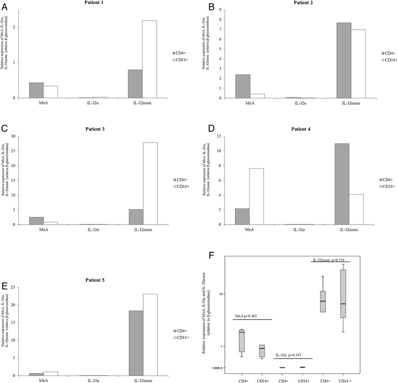
Figure 4.

mRNA expression of MxA, IL-32α and IL-32nonα in CD14+ monocytes and CD4+ T lymphocytes collected from treated HIV-1-infected patients with detectable viremia (n = 5). mRNA levels of MxA, IL-32α and IL-32nonα were analysed using real time RT-PCR assays. mRNA levels are expressed as relative expression [ΔCt method] normalized to the levels of the constitutively expressed β-glucuronidase gene. Panel A-E. Patient 1: viral load = 146 HIV RNA copies/ml; CD4+ T cell count = 450 cells/mm3. Patient 2: viral load = 80 HIV RNA copies/ml; CD4+ T cell count = 350 cells/mm3. Patient 3: viral load = 3278 HIV RNA copies/ml; CD4+ T cell count = 340 cells/mm3. Patient 4: viral load = 123,200 HIV RNA copies/ml; CD4+ T cell count = 400 cells/mm3. Patient 5: viral load = 1446 HIV RNA copies/ml; CD4+ T cell count = 895 cells/mm3. Panel F. Differences in mRNA levels between CD14+ monocytes and CD4+ T lymphocytes collected from treated HIV-1-infected patients (n = 5) were analysed using the Mann–Whitney test (CD4+ T lymphocytes vs CD14+ monocytes: MxA, p = 0.465; IL-32α, p = 0.347; IL-32nonα, p = 0.754).
