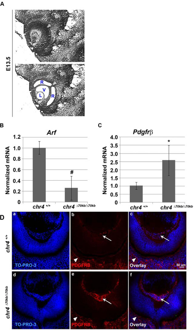
Figure 2.
Loss of the 70kb CAD risk interval decreases Arf mRNA but increases Pdgfrβ expression in vitreous of developing eye. (A). LCM of the vitreous (V), lens (L) and retina (R) from E13.5 WT mouse embryos. qRT-PCR analysis of (B) Arf and (C) Pdgfrβ were carried out using total RNA isolated from the vitreous from E13.5 chr4Δ70kb/Δ70kb embryos and wild type littermate embryos. Expression was normalized to that of Gapdh. The results were average of 3 independent experiments. # (decrease), * (increase), p<0.05. (D) Representative photomicrographs of immunofluorescence-stained sections from E13.5 embryos showing TO-PRO-3 (a, d), Pdgfrβ (b, e) and the overlay (c, f) of primary vitreous hyperplasia in chr4Δ70kb/Δ70kb embryos (d, e, f) and wild type littermates (a, b, c). Arrow (b, e) depicts Pdgfrβ staining in vitreous; arrowhead (b, e) depicts such staining in choroid/sclera.

