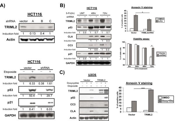
Figure 3. TRIML2 is a positive regulator of p53 activation and apoptosis.
(A) Upper panel: lentiviral transduction of control vector and three different short hairpins for TRIML2 (sh-A, -B and –C) in Hct116 cells, followed by western blot analysis for TRIML2 and loading control (Actin). Lower panel: western blot analysis for TRIML2, p53, CDKN1A (p21) and GAPDH in Hct116 cells infected with parental vector or sh-A for TRIML2. Stably-infected cells were treated with etoposide (100μM) for 24 hr. The data depicted are representative of three independent experiments. Densitometry quantification of TRIML2 levels, normalized to Actin or GAPDH, is depicted.
(B) Left panel: western blot analysis of TRIML2, p53, cleaved caspase 3 and cleaved lamin A (CC3 and CLA, both markers of apoptosis) in Hct116 cells infected with control vector or sh-A to TRIML2, treated with 5-FU (5μM) for 48 or 72 hr. Densitometry quantification of gel images was done with ImageJ software and normalized to GAPDH. The data depicted are representative of three independent experiments. Upper right panel: flow cytometric analysis of Annexin V staining in Hct116 cells stably-infected with control vector or sh-A of TRIML2, following treatment with 5-FU (5μM) for 72 hr. Bottom right panel: flow cytometric analysis of cell viability in Hct116 cells stably infected with control vector of sh-A of TRIML2, following treatment with 5-FU (5 μM) for 24, 48, or 72 hr. The averaged results from three independent experiments are shown, and error bars mark standard error. Asterisk denotes p<0.05.
(C) Left panel: western blot analysis of U2OS cells containing doxycycline-inducible vector alone (vector) or doxycycline-inducible TRIML2, following treatment with 0.75 μg/mL doxycycline and 100 μM etoposide for twenty-four hours. Cleaved lamin A (CLA) and cleaved caspase 3 (CC3) were used as markers for apoptosis. The data depicted are representative of three independent experiments. Right panel: flow cytometric analysis of Annexin V staining in U2OS cells containing doxycycline-inducible vector alone (vector) or doxycycline-inducible TRIML2, following treatment with 0.75 μg/mL doxycycline, and 100 μM etoposide for twenty-four hours. The averaged results from three independent experiments are shown, and error bars mark standard error. The triple asterisk (***) denotes p<0.0005.

