
Figure 1. ErbB2-initiatedearly lesions in p53-null mammary glands exhibit decreased senescence but intact apoptosis.
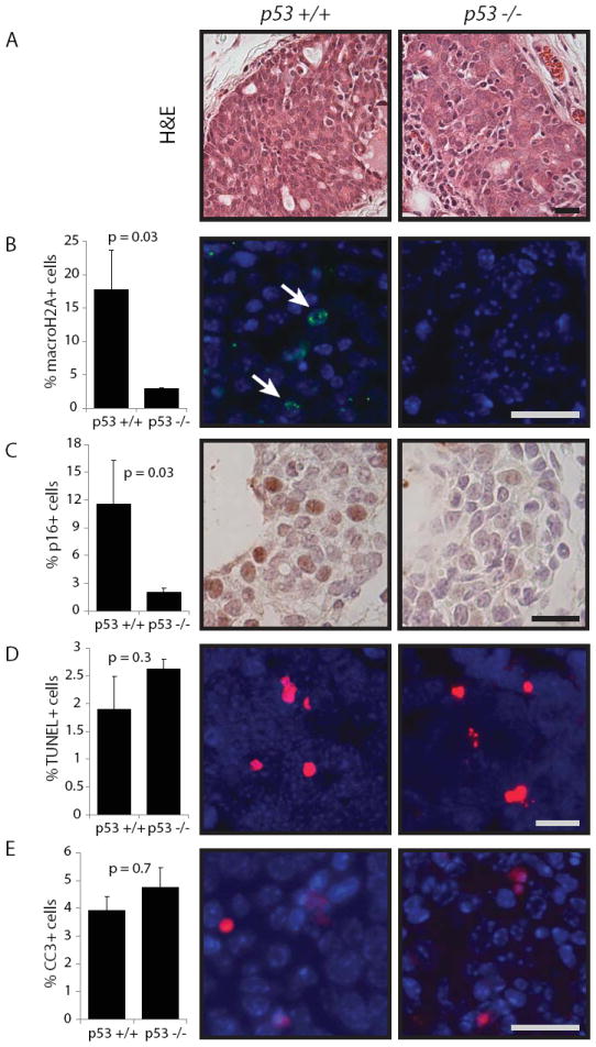
Lentivirus was used to carry caErbB2 into the mammary glands. Early lesions were analyzed two weeks following viral injection. Scale bar = 20μm. For bar graphs, columns represent the mean, and error bars represent the SEM.
A. H&E of premalignant lesions from wildtype and p53-null mammary glands.
B. Quantification of macroH2A-positive cells in premalignant lesions from widltype and p53-null mice (n=3, 4). Representative images shown.
C. Quantification of p16-positive cells in premalignant lesions from widltype and p53-null mice (n=4, 5). Representative images shown.
D. Quantification of TUNEL-positive cells (red) in premalignant lesions from widltype and p53-null mice (n=4). Representative images shown.
E. Quantification of CC3-positive cells (red) in premalignant lesions from widltype and p53-null mice (n=3,4). Representative images shown.