
Figure 4. The miR-145 Target ARF6 is overexpressed in lymph node metastasis.
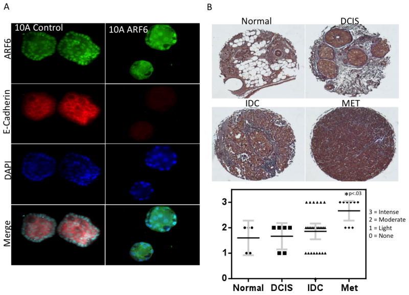
A, 3-D cell culture of MCF-10A cells stably infected with ARF6. Cells are grown in EGF supplemented media (10 ng / ml) for 7 days followed by fixation and staining with ARF6 and E-Cadherin antibodies followed by DAPI counterstaining. Acinii were examined via confocal microscopy. B, Immunohistochemistry for ARF6 was performed on a tissue microarray of matched normal and breast tumor tissue including matched primary and lymph node metastasis core samples. Normal tissue n=5, DCIS n=6, IDC n = 26, MET n = 9.