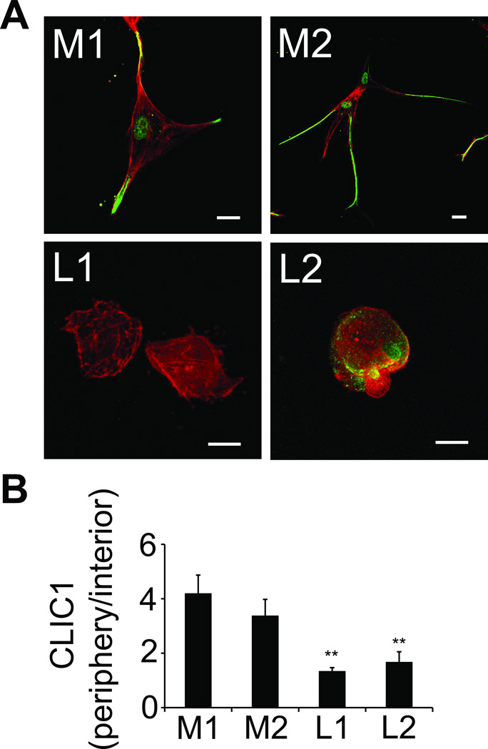
Fig. 3. CLIC1 is upregulated in invadopodia of metastatic RCC.
(A), primary kidney tumor cells from 2 patients with metastasis (M1/2) and 2 without metastasis (L1/2) were embedded in fibrin clots for 24 hours. In preparation for confocal microscopy, the fibrin-embedded cells were fixed and co-stained with anti-CLIC1 (green) and phalloidin (red). Scale bars, 20 µm. (B), CLIC1 fluorescence intensity is depicted as a ratio of peripheral to interior CLIC1 in primary tumor cells from metastatic (M) and localized (L) kidney cancer to assess CLIC1 redistribtion to invadopodia at indicated times. **P < 0.01, L1 and 2 vs. M1 and P < 0.05, L1 and 2 vs. M2.

