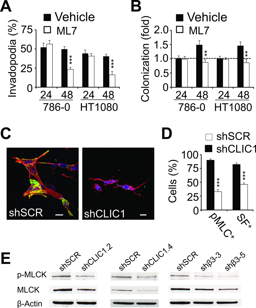
Fig. 5. CLIC1 promotes tumor cell spreading through effects on myosin-light chain kinase.
(A–B), 786-0 as well as HT1080 cells treated with 10 µM ML7 or vehicle were analyzed for invadopodia (A) and colony formation (B) 24 and 48 hours after embedding in fibrin using phase contrast microscopy. ** p<0.01, *** p<0.001. (C), confocal microscopy images of shSCR (left) and shCLIC1 (right) 786-0 embedded in fibrin (48 hours) and stained for phospho-MLC (green) as well as F-actin (red). Nuclei are stained with draq5 (blue). Scale bars, 20 µm. (D), fluorescence microscopy fields were scored for phospho-MLC- and stress fiber (SF)-positive 786-0 cells as percent of total after 48 hours embedding in fibrin. (E), extracts from 786-0 cells treated with CLIC1 and β3 shRNA (2 clones each) were probed for MLCK or phospho-MLCK. Controls were treated with scrambled shRNA (SCR). β-actin shows protein loading.

